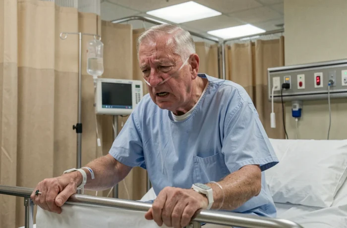
A patient arrives at the emergency room, sitting on the edge of the stretcher, gripping the rails, unable to lie down. Oxygen saturation drops, respiratory rate rises, and auscultation does not always reveal the expected crackles. Recognizing cardiogenic pulmonary edema (CPE) in its deceptive forms requires correlating several clinical indicators without waiting for a complete picture.
Wheezing and absence of crackles: the trap of cardiac asthma
One expects to hear bilateral crackles in a patient with CPE. However, in practice, this classic presentation is often absent in early forms. The stethoscope then picks up diffuse wheezing, easily confused with an asthma attack or an exacerbation of COPD.
This picture, sometimes referred to as “cardiac asthma,” is even more misleading when the patient has a history of respiratory issues. Hypoxemia and tachypnea may already be significant while crackles remain subtle or even absent. To distinguish CPE from pure respiratory decompensation, one looks for extrapulmonary elements: frank orthopnea, jugular distension, recent weight gain, history of known heart failure.
In practice, when a dyspneic patient wheezes and the bronchodilator shows no improvement in the first few minutes, it is necessary to reevaluate the cardiac hypothesis. Understanding the clinical signs of cardiogenic pulmonary edema in their variants helps avoid wasting time on a futile bronchial pathway.

Blood pressure and cardiogenic pulmonary edema: two opposing clinical profiles
Measuring blood pressure upon arrival guides towards distinct pathophysiological mechanisms, and thus towards different management strategies. Two main profiles are distinguished.
Hypertensive CPE with preserved systolic function
The patient presents with a blood pressure spike often higher than usual values, a flushed face, and agitation. Afterload suddenly increases, left ventricular filling pressures soar, and plasma fluid enters the alveoli. The ejection fraction may remain relatively normal. This profile generally responds well to nitrates and diuretics.
CPE with hypotension and signs of shock
Conversely, cold extremities, mottling, low blood pressure, and altered consciousness signal a progression towards cardiogenic shock. The cardiac pump fails, and output drops. Nitrates become counterproductive, and one moves towards inotropic support.
Taking blood pressure before any treatment conditions the subsequent management. A CPE is not just a CPE: the strategy diverges radically depending on whether the patient is hypertensive or hypotensive.
Arterial blood gas and CPE: what PaCO2 reveals about respiratory exhaustion
At the onset of CPE, reflex hyperventilation leads to normal or lowered PaCO2. The patient compensates by increasing their respiratory rate and volume. This gasometric result can falsely reassure.
The progressive rise in PaCO2 is a major alarm signal. It indicates not an aggravation of the edema itself, but an exhaustion of the respiratory muscles. The diaphragm and intercostals can no longer maintain alveolar ventilation. At this stage, the risk of respiratory arrest approaches.
In practice, one monitors the trend more than the isolated value:
- Low or normal PaCO2 with tachypnea: compensation phase, the patient is actively fighting against hypoxia
- PaCO2 starting to rise while respiratory rate remains high: ventilatory fatigue, indication to anticipate assisted ventilation
- Clearly elevated PaCO2 with bradypnea or confusion: advanced exhaustion, intubation often imminent
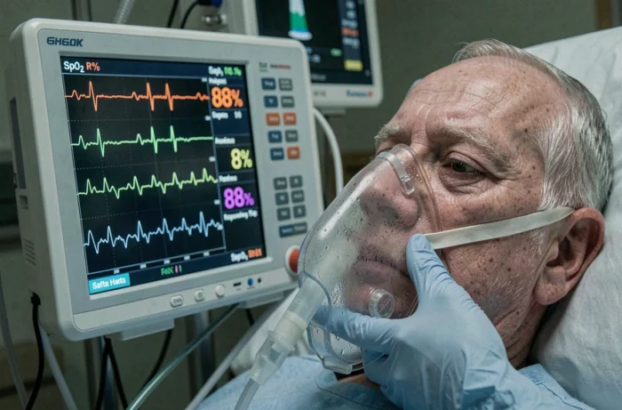
Not waiting for complete gas decompensation to consider non-invasive ventilation (NIV) with two levels of positive pressure remains a reflex to be ingrained.
Lung ultrasound at the bedside: confirming CPE without X-ray
Chest X-ray remains a reference examination for visualizing bilateral alveolar opacities in a “butterfly wing” pattern. In practice, it is not always available quickly, and its quality depends on the patient’s position (bedside image, often sitting, with sometimes unhelpful results).
Lung ultrasound performed directly at the patient’s bedside provides answers within minutes. One looks for diffuse and bilateral B-lines, these vertical hyper-echoic artifacts that start from the pleura and descend without fading. Their presence indicates an interstitial syndrome, compatible with pulmonary edema.
To strengthen the cardiogenic hypothesis, a quick assessment of the inferior vena cava is added. A dilated and poorly compliant inferior vena cava (which does not collapse or collapses little on inspiration) suggests cardiac volume overload. Combining bilateral B-lines and a dilated inferior vena cava allows for a highly probable diagnosis without waiting for an X-ray.
Feedback varies on the learning curve required, but detecting B-lines is one of the most reproducible ultrasound gestures, even for a less experienced operator.
Pink and frothy sputum: a late but specific sign of cardiogenic pulmonary edema
When the patient coughs up pink frothy sputum, the diagnosis is generally no longer in doubt. This sign indicates the passage of plasma fluid mixed with red blood cells into the alveoli, and then into the airways. Pink sputum appears in advanced forms, not at the beginning of the presentation.
Waiting for this sign to suggest CPE would delay management. Early elements (orthopnea, tachypnea, desaturation, wheezing or crackles, cardiac context) are sufficient to initiate treatment. The frothy sputum confirms retrospectively; it does not trigger the approach.
Cardiogenic pulmonary edema is rarely recognized by a single isolated sign. It is the bundle of indicators (patient position, auscultation, blood pressure, gasometry, ultrasound) that allows for swift action. The complete typical picture arrives late; treatment must begin beforehand.